The human body, at its core, is a masterpiece of biomechanics supported by 206 bones that form the skeletal framework.
These bones are not static structures but dynamic. They are constantly being remodelled and vascularized.
They provide structural support to the complex human life. Yet, malignancy can occur within this strong lattice. Osteosarcoma is the most common malignant tumor of the bone. The very regenerative mechanism is hampered, which results in local destruction of bone and distant metastasis.
Understanding the Anatomical Invasion
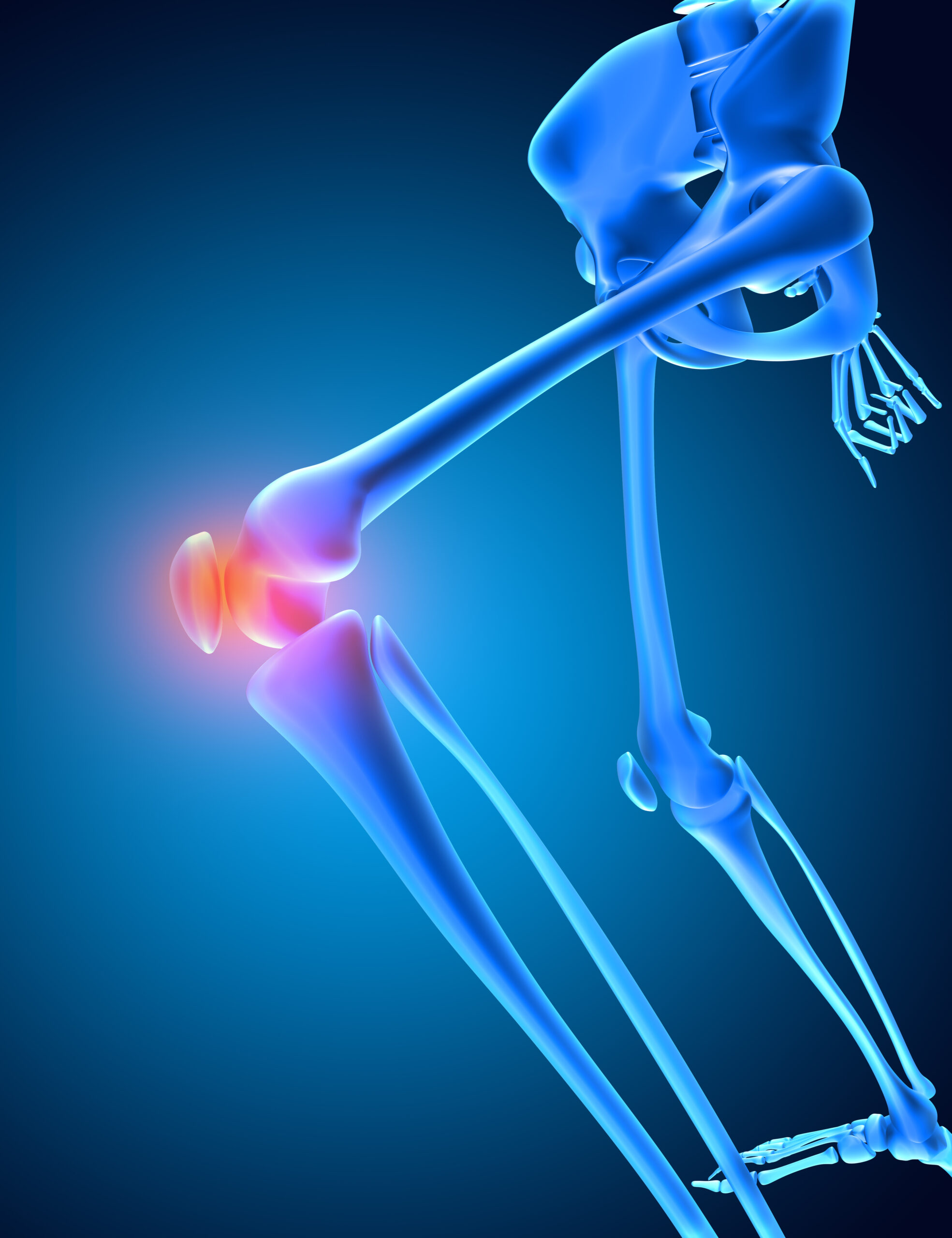
Osteosarcoma is a metaphyseal bone tumour. The metaphysis is the part of the long bone that is sandwiched between the diaphyses and the epiphyses, and is a zone of high turnover. It provides a fertile ground for cell division, resulting in malignancy. The metaphyses being highly vascular, sustain the rapidly dividing malignant cells. The haphazard osteoid matrix then invades the adjacent trabecular portion of the bone. It disrupts the normal medullary architecture of the bone and then breaches the outer strong cortical bone.
Human bone is surrounded by a fibrous membrane termed as periosteum. Periosteum functions as a barrier to the expanding tumour. It exhibits thickening. This is termed the periosteal reaction. However, if not stopped, the invasive potential of the tumour overtakes the protective periosteal reaction and utilises the rich vascularity to hematogenously spread to soft tissue and vital organs like the lungs
Epidemiology
Osteosarcoma shows bimodal age distribution. It peaks during adolescence when the growth of the long bones is maximum. A minor peak is observed among adults, which is often linked to predisposing conditions such as prior radiation exposure and Paget’s disease.
Males are slightly more affected than females. The distal end of the femur, the proximal end of the tibia, and the distal end of the humerus are common sites of osteosarcoma.
Risk factors
Most of the osteosarcoma arises de novo. However, the confluence of genetic predisposition and environmental factors plays a role in certain subsets of this malignancy.
The common risk factors identified have been highlighted below:
1) Genetic syndromes: Individuals with mutations of tumour suppressor genes like the retinoblastoma gene(RB) or TP53 have impaired ability to inhibit cellular proliferation. Such mutations can be a part of various syndromes like Li-Fraumeni syndrome (TP53 mutation), hereditary retinoblastoma (RB1 mutation), Rothmund-Thomson syndrome, Bloom syndrome, and Werner syndrome. Such individuals have an increased risk of developing Osteosarcoma.
2) Radiation exposure: Individuals with a history of exposure to radiation at high doses, especially higher than 30 Gy, have an increased risk of osteosarcoma. They can develop this malignancy even decades later.
3) Paget’s Disease: Individualswith Paget’s disease undergo constant remodelling of the bone. This predisposes the osteoblasts for malignant transformation.
4) Rapid growth of bone: There is an increased metaphyseal activity of the bone during adolescence. The rapid growth spurt in adolescence, especially in individuals with rapid longitudinal growth, is a risk factor for the development of osteosarcoma.
5) Environment and Occupational Exposure: Certain carcinogens like beryllium, vinyl chloride, and chronic bone injury might play a role in osteosarcoma.
Pathogenesis:
Osteosarcoma is characterised by instability within the genome. There is a fine balance between tumour supressors and oncogenic drivers. In osteosarcoma, the tumour suppressor genes like TP53 and RB1 are silenced. On the other hand, there is an inciting mutation that upregulates the oncogenic drivers like MDM2 and CDK4. This results in unrestrained proliferation of osteoblasts. These malignant osteoblasts secrete aberrant osteoid.
Molecular Biology
Recent advances have highlighted chemothripsis at the core of osteosarcoma. Chemothripsis involves the catastrophic rearrangement of the genes that accelerates the evolution of the tumour. There can also be improper activation of aberrant pathways like Wnt/β- catenin pathways. This results in aberrant differentiation of the osteoid matrix, thereby producing aggressive phenotypes.
The regenerative landscape of the bone is converted into a malignant ecosystem. The inflammatory cytokines and tumour-derived RANKL promote the function of osteoclasts. Osteoclasts cause resorption of bone and release of growth factors like TGF-β, which further enhance growth. Due to an increase in VEGF, neovascularisation is facilitated. This promotes metastatic escape routes. The perfusion is often inadequate to meet the demand of rapidly dividing tumour cells. This creates a hypoxic acidic niche that promotes the selection of aggressive tumour clones and facilitates drug resistance.
Clinical Presentation: Pain that whispers before it screams
The onset of osteosarcoma is insidious. There is a dull localised bone pain. This is followed by a swelling and a functional limitation. As the tumour disrupts the cortex, there can be visible masses due to soft tissue involvement and pathological fractures. Systemic symptoms are present in metastasis.
Radiological Hallmarks
The typical radiographical findings in osteosatcoma can be attributed to the periosteal reaction. It reveals a mixed lesion with ill-defined margin. Periosteal reaction is visible as Codman’s triangle and sunburst patter. MRI is useful to rule out soft tissue involvement, while CT scan helps in assessing pulmonary metastases.
Histopathology
Under a microscope, osteosarcoma can be diagnosed if there is presence of large number of osteoid producing malignant osteoblasts. Typical neoplastic changes can be seen within the cell which includes atypical mitosis, necrosis, increased nuclear to cytoplasmic ratio, pleomorphism, etc.
Staging and Prognosis
The Enneking and AJCC systems are used for staging osteosarcoma. It incorporates tumour stage, local extent, and presence of metastases. The prognosis depends upon the surgical margins, response to the neoadjuvant chemotherapy and presence of metastasis at presentation. If there is involvement of lung, the prognosis is poor and the five year survival rate decreases to 30 %
Treatment
Just as any other neoplasm, osteosarcoma warrants a multimodal approach. The tripartite approach includes, surgical resection, neoadjuvant and adjuvant chemotherapy.
Surgery
Current advances allow limb sparing surgery. In case of tumours involving neurovascular structure, amputation remain surgery of choice. The adequacy of surgery requires complete resection of tumour with negative margins.
Chemotherapy
The standard regimen includes high dose methotrexate, doxorubicin, cisplatin. These drugs play a pivotal role in achieving micrometastatic control.
Survivorship and Late Effects
Survivors often live with the long-term complications of chemotherapy. There can be variable functional outcomes following a limb surgery or an amputation. Care must address both biological and emotional sequelae of the disease.
Conclusion
Osteosarcoma creates a genetically unstable, structurally disfigured and biologically hyperactive scenario. The chaotic proliferation of osteoid warrants scientific audacity and surgical precision. The development of immunotherapy has added new weapons in our arsenal for the treatment of osteosarcoma. Genetic counselling and surveillance is crucial, especially in high-risk populations. Judicious use of therapeutic radiation, earlier detection of symptoms in Paget’s disease, are some of the definitive prevention strategies for Osteosarcoma. Researchers, through their conceptual understanding of mechanisms, can transform this into proactive intervention steps to provide newer wings of hope for the management of osteosarcoma.
References:
- Mirabello L, Troisi RJ, Savage SA. Osteosarcoma incidence and survival rates from 1973 to 2004: data from the Surveillance, Epidemiology, and End Results Program. Cancer. 2009 Apr 1;115(7):1531–43.
- Kansara M, Teng MW, Smyth MJ, Thomas DM. Translational biology of osteosarcoma. Nat Rev Cancer. 2014 ov;14(11):722–35.
- Ottaviani G, Jaffe N. The epidemiology of osteosarcoma. In: Jaffe N, Bruland OS, Bielack S, editors. Pediatric and Adolescent Osteosarcoma. Boston, MA: Springer; 2010. p. 3–13.
- Luetke A, Meyers PA, Lewis I, Juergens H. Osteosarcoma treatment—where do we stand? A state of the art review. Cancer Treat Rev. 2014 Jan;40(4):523–32.
- Isakoff MS, Bielack SS, Meltzer P, Gorlick R. Osteosarcoma: Current treatment and a collaborative pathway to success. J Clin Oncol. 2015 Feb 20;33(27):3029–35.