Introduction
Tobacco smoking is one of the biggest global epidemics, killing almost half of its users who don’t quit. Smoking predisposes individuals to chronic obstructive pulmonary disorder or COPD, which results in functional or anatomical obstruction of small airways. Functional obstruction is manifested as Emphysema while anatomical obstruction manifests as chronic bronchitis.
COPD is the 4th leading cause of death worldwide. The burden is especially high in lower and middle-income countries, which account for almost 90% of the deaths due to COPD. This is attributed to rampant indoor air pollution and smoking.
Emphysema is characterised by progressive irreversible destruction of the alveoli. Despite its prevalence, it is still termed as a silent epidemic as many cases remain undiagnosed until advanced stages. Through this article, we try to understand the mechanism of emphysema and its clinical features, as well as explore the available treatment options.
Biology of breathing
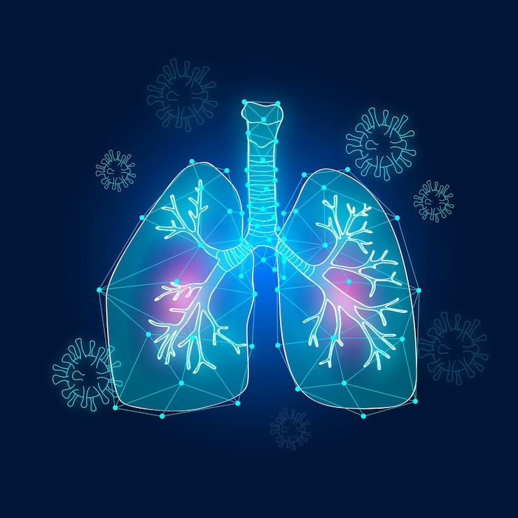
We need O2 to survive. The presence of a pair of lungs assists in delivering oxygen while filtering out carbon dioxide. During inhalation, the air travels into the trachea, facilitated by the pressure gradient. It makes its way through the airways into the tiny balloon-like air sacs known as the functional unit of the lungs: the alveoli. The membrane of the alveoli serves a vital role in diffusing oxygen into the blood vessels. They expand on inspiration and recoil during expiration. The elasticity of the alveolar wall is crucial in this function. This elasticity is maintained by an alveolar membrane protein called elastin.
Chronic exposure to toxic substances like cigarette smoke, industrial pollutants, etc, leads to the destruction of elastin protein. This permanently expands the alveoli and reduces the surface area for oxygen diffusion. It leads to chronic breathlessness, air trapping and hyperinflation of the lungs
Pathophysiology
Emphysema involves a complex interplay of oxidative stress, chronic inflammation, protease-antiprotease mismatch and toxic injury.
- Toxic Injury: Exposure to toxins like cigarette smoke, industrial pollutants triggers inflammatory mediators like macrophages, neutrophils. These cells release cytokines like TNF-alpha, IL-8 that result in cellular damage, tissue remodelling and parenchymal destruction.
- Oxidative stress: Reactive oxygen species released during inflammation incur irreversible damage by free radical mechanism. The oxidants are usually kept in check by potent antioxidants like SOD3. However, the transcription factor NRF2, a major regulator of oxidant-antioxidant balance, is upregulated, resulting in increased production of matrix metalloprotease-like macrophage elastase.
- Protease: Antiprotease imbalance: An enzyme α1 – antitrypsin is a potent antiprotease and inhibits the degradation of elastin protein by elastases. Genetic deficiency of the enzyme predisposes individuals to emphysema.Recent guideline have made testing for α1 – antitrypsin imperative for all subjects of COPD.
- Infections: Infections triggering inflammation remain an independent risk factor for emphysema. Adult respiratory infections result in exacerbations of COPD episodes.
The disease is characterized by a reduction in forced expiratory flow rates, unresponsive to bronchodilators. There is dilatation of airways resulting in air trapping and an increase in the residual volume of the lungs, The lungs are hyperinflated and voluminous.
Types of Emphysema
The anatomical classification of emphysema is based on the site of dilatation of acini. An acini is made of an alveolar sac, an alveolar duct and a terminal respiratory bronchiole. Emphysema can be Centriacinar (dilatation of the terminal respiratory bronchiole), Panacinar (regular dilatation of the entire acini), Paraseptal (dilatation of the distal airways, along the pleural septae) and Irregular (irregular dilatation of the acini). Centriacinar is the most common type and is associated with smoking. Pan acinar is associated with α1-anti-trypsin deficiency. Paraseptal usually occurs in younger populations and results in spontaneous pneumothorax.
Clinical Signs and Symptoms
Difficulty in breathing or dyspnea, is the first symptom. The patient has a barrel-shaped chest, in a hunched-over position. There is visible hyperventilation. This is a compensatory mechanism that allows CO2 washout and prevents cyanosis. Because of the relatively normal blood gas values, these patients are termed as pink puffers. The persistant hypoxia in the advanced cases causes damage to the pulmonary vessels, resulting in pulmonary failure, coma, or right sided heart failure.
Establishing the diagnosis
Earlier detection of emphysema is crucial for preventing complications. The primary investigation to characterize the disease is spirometry. The patient is instructed to inhale air to the maximum capacity. The patient then exhales and the volume of air exhaled during first second (FEV1) and the total volume of air exhaled during entire spirometry procedure (FVC) is measured. The ratio of FEV1/FVC is reduced in patients with COPD. Based on these values, the severity of the disease is assessed. The criteria used is Golds Criteria given by Global Initiative for Chronic Obstructive Lung Disease.Stage I and II have little to no emphysema. Stage 3 and Stage 4 are advanced stages demonstrating extensive emphysema.
GOLD Criteria:
- Stage I : Mild severity : FEV1 /FVC <0.7 and FEV1 ≥80% predicted
- Stage II: Moderate severity: FEV1/FVC <0.7 and FEV1 ≥50% but <80% predicted
- Stage III: Severe: FEV1/FVC <0.7 and FEV1≥30% but <50% predicted
- Stage IV: Very severe: FEV1/FVC <0.7 and FEV1<30% predicted
Assessment of the diffusion capacity of Carbon monoxide across the alveolar membrane is a diagnostic method to check its efficiency. The diffusion is increased due to increased capillaries, owing to inflammation. High resolution CT scan is the gold standard investigation, assisting to visualize the alveolar damage.
Treatment
The functional obstruction in Emphysema can only be symptomatically management. Treatment mainly focuses on slowing the progression of the disease. Airflow can be improved by administration of Bronchodilators. The inflammation is reduced by inhaled corticosteroids. Genetic therapy is recommended in enzyme deficiency cases.
The only modality to prevent further damage is smoking cessation. Exercise training increases the efficiency of oxygen utilization and reduces dyspnea. Long volume reduction surgery is used in advanced cases. The last resort is to replace the irreversible damage by lung transplantation.
Conclusion
Emphysema is a preventable disease, yet it affects millions worldwide. The best way to manage this devastating condition is by never smoking, reducing pollution exposure by adopting strategies like wearing masks in poor air quality index places. It is imperative to prioritize lung health from an early age to prevent becoming a victim. Early detection, portable spirometry camps, smoking cessation campaigns can reduce the burden as prevention is the best cure for COPD.
References:
1. Barnes PJ. Inflammatory mechanisms in patients with chronic obstructive pulmonary disease. J Allergy Clin Immunol. 2020;145(1):16-27. doi:10.1016/j.jaci.2019.11.010
2. Shapiro SD. Proteolysis in the lung. Eur Respir J Suppl. 2003;44:30s-32s. doi:10.1183/09031936.03.00000403a
3. MacNee W. Pathophysiology of cor pulmonale in chronic obstructive pulmonary disease. Am J Respir Crit Care Med. 1994;150(3):833-852. doi:10.1164/ajrccm.150.3.8087332
4. Global Initiative for Chronic Obstructive Lung Disease (GOLD). Global Strategy for the Diagnosis, Management, and Prevention of COPD: 2023 Report. Published 2023. Accessed March 30, 2025. https://goldcopd.org
5. Chambers DC, Enever D, Ilic N, et al. Mesenchymal stromal cell therapy for chronic lung disease: Reshaping the horizon. Respir Res. 2022;23(1):50. doi:10.1186/s12931-022-01942-7